March 19, 2018
Hugo Sinha, a master’s student in the Department of Electrical and Computer Engineering at Concordia University in Montreal, Canada, is hoping to make automated CRISPR technology accessible to as many cancer researchers as possible. His vision is to “empower all cancer biologists to be able to program their gene editing experiments on their own benchtop instruments.” As one of the youngest presenters at SLAS2018 and an SLAS2018 Tony B. Academic Travel Award winner, Sinha presented his work in a podium presentation “Automating Gene Editing for Deciphering Cancer Pathways Using Microfluidics.”
Sinha makes the case for automating CRISPR-Cas9 (CRISPR) workflows on an affordable platform. “In cancer research, dealing with complex molecular pathways and implicating numerous gene suspects, researchers use gene editing in an iterative process and it is that fundamental aspect (many iterations) that is problematic,” he explains. “The issue is that we are looking for genes in a haystack, often addressed with automated robotics that can increase throughput but are burdened with high cost and expertise requirements, leaving smaller labs and start-ups to use manual interventions that are slow and prone to human error.” Sinha saw these limitations as inspiration to develop a miniaturized automated platform tailored to gene editing pipelines.
As presented at SLAS2018, highlights of his digital microfluidics system include:
“CRISPR is the most popular technique in use today for gene editing due to the ease of customizing it to many tasks,” Sinha says. “Its broad applicability has produced a recent surge in publications with no signs of slowing down.” A PubMed search quickly reveals the truth in that statement. Furthermore, in a recent paper reviewing applications of CRISPR in cancer biology, the authors conclude “Ever since the Doudna and Charpentier groups demonstrated the potential of the CRISPR-Cas9 system as a powerful RNA-programmed gene editing platform [2012], the field of genome engineering has rapidly undergone a scientific revolution that promises to transform nearly every aspect of basic biological and biomedical research.” In fact, Sinha pointed out a paper published this January, that reports on how researchers “have modified the Cas9 enzyme (named xCas9) to make it more specific compared with the standard Cas9 enzyme.”
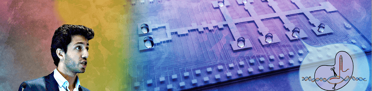
“CRISPR works like a pair of scissors to cut, insert or reorder specific genetic fragments, creating changes in the biological cell that can then be studied to better understand gene function,” Sinha explains. “Scientists can target gene suspects in metabolic pathways, alter them and evaluate the resultant phenotype arising from genomic disruption.” Some of the benefits and challenges of CRISPR are reviewed in this SLAS paper in which the author notes the system “has now been widely adopted by the entire life science community, and novel applications of the CRISPR systems are appearing at a breakneck pace.”
According to Sinha, the automation equipment developed in the Shih Microfluidics Laboratory at Concordia costs less than $10,000 and is comprised of readily available components. One core piece of the system is an Arduino microcontroller that he describes as an open-source electronics platform that makes electrical engineering accessible to anyone with a DIY mentality. Due to the complexity of the programming for this project, the software was designed by Philippe Vo, an engineer in Shih’s lab. The automation is publicly available on GitHub because Sinha and Shih want to make the technology available to as many researchers as possible. The open-source movement is at work in a number of fields and last June, Metafluidics, an open-source, community-driven repository for microfluidics was started.
Sinha reports success on a proof of concept, miniaturized, automated platform tailored to the CRISPR gene editing workflow that includes all steps from initial seeding of cells to capturing the experimental results at the end. He considers “the ability to carry out all the steps of the workflow without having to transfer to another platform a key piece in making the device successful.”
“Microfluidics is part of the larger realm of miniaturization where you integrate lab processes onto a tiny handheld device and it can be approached in several ways,” Sinha explains. Two approaches use fixed, enclosed micron-sized channels to move fluid either in a continuous stream into chambers or as discrete droplets that act as individual test tubes. The third approach is called digital microfluidics (DMF).
DMF is a “liquid-handling technology that enables individual control over droplets on an open array of electrodes. These picoliter- to microliter-sized droplets, each serving as an isolated vessel for chemical processes, can be made to move, merge, split, and dispense from reservoirs,” state the authors of a comprehensive review paper. They note that DMF has been used in a large number of applications in a wide range of fields which involve chemical and enzymatic reactions, immunoassays, DNA-based applications, clinical diagnostics, proteomics and cell-based applications.
What Sinha likes about DMF is that “you can perform all the operations you do with liquids at the bench (dispensing, moving, merging, mixing and splitting) and put them on the chip format. The direct translation of workflow from bench to chip makes the processes feel familiar even though fluid is manipulated as discrete droplets on an open array of electrodes. Compared with channel-based systems, this approach has the advantages of active mixing (rather than passive diffusion), of addressing samples individually (eliminating cross-contamination), of integration with detectors for analysis (e.g. fluorescence or absorbance) and of automation.”
There is a lot of detail in the engineering and the design of the chip itself but Sinha explains that “most of the components and processes are fairly standard and then the biology and chemistry just need to be optimized as it would in any format.” With the goal of creating something that would be used in a biologist’s laboratory, Sinha has put a lot of thought into how to make the device intuitive for use. The team designed a working model of a graphical user interface so that users could upload their device design, set up the droplet parameters, upload the droplet sequence, monitor progress in real time through a viewing window and get feedback. The image-based feedback system, developed in the lab (and published last year), is really important for success, he says. It was developed to make sure that the droplets are moving as they should, and if not, a series of operations occurs to ensure the droplet progresses.
With a firm understanding of the engineering behind the chip, Sinha set out to determine if the biology would work. He explains there were four key processes to evaluate on the device: establish healthy cell cultures, transfect cells efficiently, use Cas9 to knock out the test gene, and finally, to validate the device for cancer research by targeting oncogenes that are involved in the cell proliferation pathway.
“I wanted to make an intuitive design, arranging the electrodes in a specific way that would simplify operations on device to ensure the cells are as healthy as possible for as long as possible,” he describes. “I wanted the whole gene-editing pipeline to be automated on this single device—to load cells onto the chip at the beginning and to analyze the results in about seven days. Usually with robotics or other platforms using microfluidics, there is a point where you have to get the cells out of the device to perform some steps. I wanted to avoid that.”
Since the goal was to use the chip with CRISPR for cancer research, the team selected a non-small cell lung carcinoma cell line (H1299) with stable expression of enhanced green fluorescent protein (eGFP). Sinha demonstrated that cells grown on the device were not morphologically different from those grown in a standard 24-well plate. Therefore, the design of the lab-on-a-chip was functional. Next Sinha, validated the system by transfecting a red fluorescent gene into the cells. He describes the process of developing a system to quantitatively evaluate transfection efficiency while leaving the cells undisturbed on the chip. He explains that the goal of on-chip analysis meant that “we were keen to use microscopy and had to develop an approach that would allow accurate counting for both transfection and later knock-out efficiency. The 3D stacking of cells makes this a challenge, but using a cell profiler program and nuclear stains, the positive cells could be counted accurately.”
Optimizing the transfection efficiency involved some additional work, to get the right lipid complex:media ratio, and eventually achieve transfection rates just slightly less than those in 24-well plates. With the transfection protocol developed, Sinha tested the device with the CRISPR system to knock out the GFP gene expressed in the selected cell line. This worked well and the last step is to prove that the system can be used to edit cell proliferation pathways in these cancer cells. Sinha chose the ras protein kinase cascade to test the assay and is working in 24-well plates to optimize experimental conditions. On-device work is to be completed in the next few months but Sinha is confident that the biology and engineering will work well together.
“We have developed a fully automated transfection device, optimized transfection conditions on the device, used the device for CRISPR-Cas9 knock-out targeting stably integrated eGFP in a non-small cell lung carcinoma line and are in the process of validating the platform for cancer research by targeting proliferation genes,” Sinha summarizes.
Sinha’s platform was well received at SLAS2018 and sparked questions from the audience. When asked about the throughput rate of the device Sinha explained that his approach “is less about absolute throughput on a single device and more about increasing access to automated assays so that more people can be working on their ideas, and that collectively, there is a greater amount of work being done.”
Sinha is finishing up some experimental work to strengthen the story and is planning to submit his work for publication. When that is completed, Sinha says “a few of us from the lab are starting a company with Shih based on digital fluidics applications. As a versatile and reprogrammable liquid handling platform there are a wide range of applications that could be developed. We have to decide what our niche will be.”
Prior to joining the Shih lab, Sinha earned his biology degree at Concordia University, were he had gravitated toward “courses related to the synthetic biology framework and was fascinated with the potential of gene editing technology enabled by CRISPR systems.” As he investigated options for graduate studies, Sinha met Steve Shih, Ph.D., an expert in microfluidic technology who also had an interest in automating synthetic biology processes. Shih was just starting his interdisciplinary lab based on his belief that "we need to bring engineers and biologists together to solve major biological and health issues.” Both Sinha and Shih were interested in exploring how synthetic biology workflows, such as CRISPR-Cas9, might be adapted to a microfluidic system and applied to cancer research. Sinha, excited about the opportunity to do research using biological and engineering technologies and to be part of a start-up laboratory, was the first student to join the Shih Microfluidics Laboratory.
In only a couple of years, Shih’s lab has grown to approximately 10 graduate students. Sinha says “it's an interesting lab because we have people who are either primarily engineers (mostly electrical or computer) or from the life sciences. Individual projects are highly collaborative. As a biologist, my project is predominantly biological. I am trying to prove that the engineering works with the biology and the opposite is true for the engineers. We are part of a larger center called The Centre for Applied Synthetic Biology. And that's exactly why Steve [Shih] suggested I go to SLAS2018. My work is in line with the SLAS mission and desire to bridge the gap between automation and life sciences.”
SLAS2018 was the first society conference that Sinha has attended. He was “very impressed at the caliber of the scientific knowledge and presentations and honored to receive a very generous SLAS2018 Tony B. Academic Travel Award.” Tony B. Award applications for SLAS2019 are due late summer 2018 and are awarded in the fall after evaluation by a selection committee. Students, graduate students, post-doctoral associates and junior faculty (less than four years in first academic appointment) may apply. The applicant must be the primary author of a submitted abstract and must present their research in either a poster or podium presentation at the conference. Those selected receive complimentary travel, lodging and registration to participate in SLAS2019.
High-Throughput Silencing Using the CRISPR-Cas9 System: A Review of the Benefits and Challenges
Droplet-Based Microfluidics: Enabling Impact on Drug Discovery
Digital Microfluidics for Automated Hanging Drop Cell Spheroid Culture
New Research: Automated Gene Editing to Detect Cancer 'Suspects'
Lab on a Chip. Concordia University Podcast, Episode 12, Release Date: November 24, 2017
Open-Source, Community-Driven Microfluidics with Metafluidics
Discovery of Cancer Drug Targets by CRISPR-Cas9 Screening of Protein Domains
Applications of the CRISPR-Cas9 System in Cancer Biology
CRISPR-Cas9 Knockin Mice for Genome Editing and Cancer Modeling