October 14, 2019
A special issue of SLAS Technology showcases the technology demands for personalized medicine and reveals how life sciences engineers approach the development cycle with the end user in mind to ensure translational opportunities. “Many times you create an innovative instrument that works perfectly in your lab and never really gets translated into use where it’s most needed,” says Claire Hur, Ph.D. “We need to make tools and techniques that the clinical technician or the patient can use – not just the Ph.D. student who developed it.”
She is excited to see this goal shared by other researchers featured in a new SLAS Technology Special Issue on Engineering Innovations for Fundamental Biology and Translational Medicine. “I like that there are engineers who are thinking about integrating their innovative technology into clinical workflow,” says Hur, who serves as co-guest editor of the special issue along with Deok-Ho Kim, Ph.D., a faculty member in the Department of Biomedical Engineering and Department of Medicine at Johns Hopkins University School of Medicine.
“All the papers in this special issue address how to streamline technological innovations for diagnosis and prognosis so that when the medical team finds the correct solution for the patient, they can implement treatment more quickly,” says Hur. She holds this standard over her own work as an assistant professor in the Department of Mechanical Engineering at the Whiting School of Engineering, Johns Hopkins University (Baltimore, MD, USA). The microfluidics expert studies individual response to different therapies and is developing liquid biopsies to get the most information possible from patient-derived biospecimens, specifically blood.
“After finding circulating tumor cells (CTCs), I am expanding my research into immune cells so we can use a patient’s specimens to quickly gather accurate information and find less toxic therapy,” says Hur. “I have two pillars of my work: One, to get the CTCs from the blood while they’re alive for assays, and two, to train the immune systems of the patients so that we can give them safe treatment. For these pillars, I am making instruments that can be used automatically and easily without bringing in a lot of highly skilled research technicians.”
Hur hopes that the special issue will reach technology engineers and influence them as they begin creating new systems to examine the entire development loop, one with the end user in mind. “I would like it if they could make adjustments as the tool or technique evolves and see consequences before they happen,” Hur explains. “When the technique comes out, it should be ready to use. It shouldn’t take another 20 years for it to be fine-tuned.”
Predictive, preventative, personalized and participatory – of the four Ps of personalized medicine, Hur comments that it is the “participatory” factor that is often overlooked as innovations evolve. She adds that the four original research articles in the special issue minimize that hurdle.
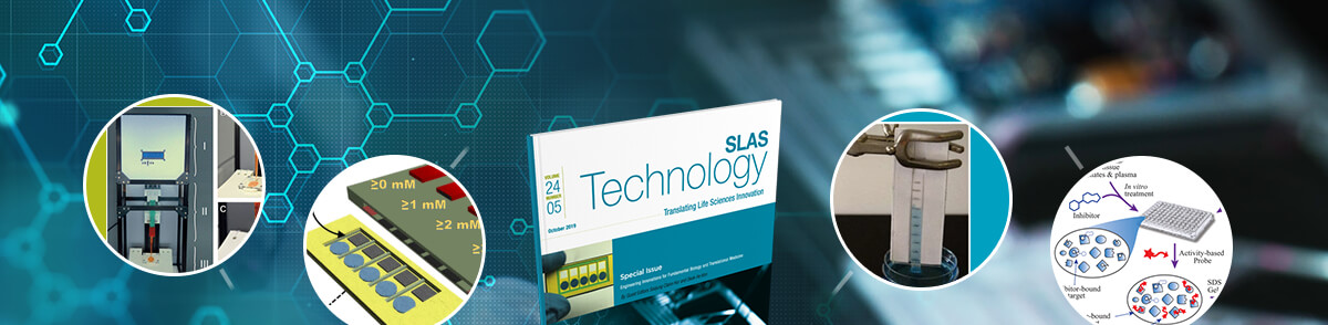
In “An Equipment-Free, Paper-Based Electrochemical Sensor for Visual Monitoring of Glucose Levels in Urine,” Maedeh Mohammadifar, Ph.D., and fellow authors from the Bioelectronics and Microsystems Laboratory of the Department of Electrical & Computer Engineering, State University of New York (SUNY; Binghamton, NY, USA), give careful attention to end-user participation by creating a noninvasive glucose biosensor that integrates a disposable, paper-based sensing strip and a simple amplifier circuit with a visual readout. The paper strip consists of five enzyme-activated electrodes that connect to a specific indicator circuit that triggers a light-emitting diode (LED) when a predefined glucose concentration is reached.
“The mechanism is as straightforward as possible, but also very cheap and disposable, ensuring that patients will succeed in using it. Their device promotes patient control of their own health monitoring,” Hur states. “While the sensor is simple, it presents a semiquantitative visual screening of specific glucose concentrations in urine that minimizes user error.”
Another example of thoughtful innovation is found in the article, “Quantification of In Vivo Target Engagement Using Microfluidic Activity Based Protein Profiling,” from Holly T. Reardon, Ph.D., and fellow authors from Abide Therapeutics (San Diego, CA, USA) and Janssen Research and Development (Spring House, PA, USA).
“The team from Abide ensure that their method can be used effectively in the clinic by incorporating it into the clinical workflow for quicker, more accurate assays,” says Hur. She describes how researchers adapt protocols for a commercial LabChip GXII microfluidic instrument to permit electrophoretic separation of probe-labeled proteins in tissue lysates and plasma and quantification of fluorescence (probe/protein labeling ratio of 1:1). The method accelerates workflows and avoids imaging artifacts that make conventional gels challenging to quantify. The time savings allows the incorporation of microfluidic activity-based protein profiling early in the drug discovery workflow, enabling routine assessments of tissue distribution and engagement of targets and off-targets in vivo.
In “Fabrication of Bioengineered Skin by Injection Molding: A Feasibility Study on Automation,” S. Fox, Ph.D., and fellow authors explore the rapid, low-cost fabrication of standardized bioengineered skin. Hur notes that “instead of using lab grown tissue, the research team produces larger scale skin for healing wounds of individual patients in the clinic by using an injection molding technique from the manufacturing field.”
According to the authors from the Product Development Group Zürich, Department of Mechanical and Process Engineering, ETH Zürich (Zürich, Switzerland), the use of bioengineered skin facilitates fundamental and applied research because it enables the investigation of complex interactions between various cell types as well as the extracellular matrix. The authors explain that the quality, standardization and production volume of living tissues under manual fabrication is dependent on the technician’s experience. To expand the quality, quantity and potential use of living tissues to a greater number of research groups, the team uses a time- and money-saving automation technique to form tissue by injecting dermal and epidermal skin layers into a customized mold. This allows the researchers to demonstrate the biocompatibility of this fabrication process and confirm the resulting bilayered morphology of the bioengineered skin using histology and immunohistochemistry.
Hur comments that another article from the special issue, “Controlling Macroscopic Phase Separation of Aqueous Two-Phase Polymer Systems in Porous Media,” shows “the researchers separating analytes from biosolution without using harsh chemicals that can damage the biomarker,” she says. “The researchers test a new way of using chemical property so that they can preserve the biomarker’s activity and do the test quicker. This procedure could be accomplished in a resource-limited setting.”
In the article, David Y. Pereira, Ph.D., and fellow authors from University of California at Los Angeles’ (UCLA) Department of Bioengineering and the Division of Advanced Prosthodontics at the Weintraub Center for Reconstructive Biotechnology, School of Dentistry (Los Angeles, CA, USA), explore a unique phenomenon from previous work in which a mixed polymer–salt or mixed micellar aqueous two-phase system (ATPS) separates into its two constituent phases as it flows within paper.
The authors note that while these ATPSs work well in their respective studies to concentrate the target biomarker and improve the sensitivity of the lateral-flow immunoassay, different ATPSs can be advantageous for new applications based on factors such as biomarker partitioning or biochemical compatibility between ATPSs and sample components. However, since the mechanism of phase separation in porous media is not completely understood, introducing other ATPSs to paper is an unpredictable process that relies on trial and error experiments. This is especially true for polymer–polymer ATPSs in which the characteristics of the two phases appear quite similar.
This led the researchers to develop semiquantitative guidelines for choosing ATPSs that can phase separate in paper. Their latest research successfully demonstrates the phase separation phenomenon in hydrogels that extend its application and potential benefits to an alternative porous medium.
The other two articles in the special issue are comprehensive review papers that Hur describes as thoughtful exploration of the technology demands in personalized medicine. “The review articles offer a comprehensive outlook of where this field is going and what is still needed,” Hur comments. “The authors emphasize that creating the right model, getting the right specimen from the patient and the importance of it help the medical team understand exactly what’s going on with the patients.”
For example, in “Biomicrofluidic Systems for Hematologic Cancer Research and Clinical Applications,” researchers examine microfluidics cell-based technology with application toward studying hematologic tumor microenvironments (TMEs) for the purpose of drug discovery and clinical treatment selection.
Mosfera A. Chowdury, Ph.D., and fellow authors from the University of Toronto’s Department of Mechanical & Industrial Engineering and Institute of Biomaterials & Biomedical Engineering, along with the Toronto General Hospital Research Institute, University Health Network, (Toronto, Ontario, Canada), discuss the persistent challenge in developing personalized treatments for hematologic cancers – namely the lack of patient specific, physiologically relevant disease models to test investigational drugs in clinical trials and to select therapies in a clinical setting.
The team provides an overview of state-of-the-art microfluidic systems designed to address questions related to hematologic TMEs and drug development and reviews pharmaceutical drugs and different modes of immunotherapy for hematologic cancers, followed by key considerations for developing a physiologically relevant microfluidic companion diagnostic tool for mimicking different hematologic TMEs for testing with different drugs in clinical trials.
In the review article, “Advances in Technologies for Purification and Enrichment of Extracellular Vesicles,” Pan Zhang and fellow authors from the National University of Singapore’s (NUS) Graduate School for Integrative Sciences & Engineering and Institute for Health Innovation & Technology (Singapore), introduce technique advances in isolating microvesicles and exosomes according to approach. The review also covers the limitations of currently reported technologies in terms of their specificity and efficiency, and provides the authors’ thoughts about future developments of extracellular vesicles (EVs) purification and enrichment technology.
The review authors report that despite the wide interest in EVs, the technologies for the purification and enrichment of EVs are still in their infancy. The isolation of EVs, especially exosomes, is inherently challenging due to their small size and heterogeneity.
In addition to this, the special issue also includes an auto commentary, "Framework Nucleic Acids: A Paradigm Shift in Transdermal Drug Delivery," with authors from Nanyang Technological University's School of Chemical and Biomedical Engineering and Northwestern Institute for Nanomedicine (Singapore). The commentary highlights the paradigm shift of nucleic acid delivery from being a cargo moiety to serving instead as a drug carrier. Author Christian Wiraja and fellow authors also discuss further development directions to maximize the potential of framework nucleic acids for transdermal drug delivery.
“For personalized medicine to be effective, we need to focus on automating all aspects of these tools and technologies to minimize user error and overcome the rate of disease progression,” Hur says. She expands on this thought in the editorial for the special issue, commenting that the growing understanding of genomics and increasing clinical evidence reveal that prescribing treatments based on population average is not an effective healthcare strategy.
“We don’t know who will develop resistance to treatment or why some have better prognoses than others,” Hur says. “I hope to expand more into this research with my own work in the future. I want to contribute to finding the correct individual patient information so we can quickly deliver the best therapy possible so that the patient doesn’t suffer from all the adverse consequences of treatment with the wrong drug for his or her cancer.”
The SLAS Technology Special Issue is available now at SLAS Technology Online for SLAS members, SLAS Technology subscribers and pay-per-view readers. Free public access becomes available one year after final (print) publication.
Join Johns Hopkin’s Claire Hur, Ph.D., as she talks about the SLAS Technology Special Issue on Engineering Innovations for Fundamental Biology and Translational Medicine in a 2019 SLAS Technology podcast.
In the News: NIH Donates $29.5M to Advance Genomics, Precision Medicine